
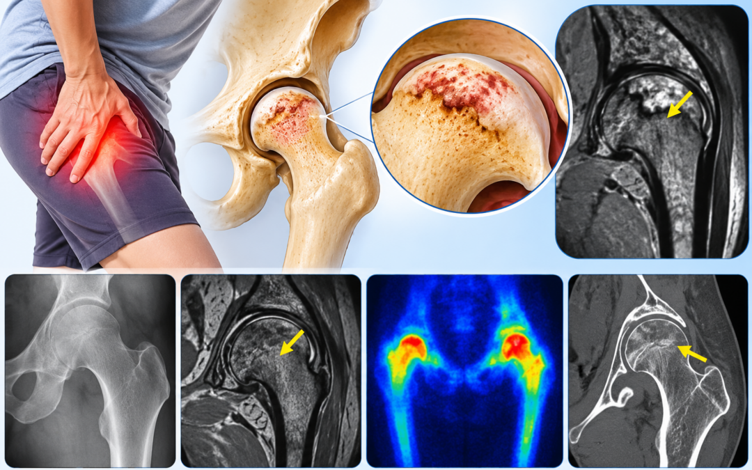
Avascular necrosis (AVN), or osteonecrosis, is what happens when blood stops reaching a section of the bone. The cells there die off and the bone slowly weakens. It can end in collapse. The hip is the usual victim. What makes it hard to catch is the start: a dull ache, sometimes nothing else, and an X-ray that looks perfectly normal. Imaging for avascular necrosis, chosen well, can pick it up before the bone gives way.
According to Dr. Gayana Shankaramurthy, a Nuclear Medicine Specialist in Bangalore at KIMS Hospitals, many patients are told for months that their X-ray is fine. “A normal X-ray doesn’t rule it out,” she says. “If the pain is persistent and the risk factors are there, further imaging is worth doing early.”
Which Tests Are Used for Avascular Necrosis Imaging?
Doctors rarely jump straight to the most advanced scan. They begin with the simplest and move up only if the picture doesn’t add up.
|
Test |
What it shows |
Where it fits |
|
X-ray |
Bone shape and collapse |
First test. Often normal in early disease |
|
MRI |
Changes in bone marrow |
Most sensitive for early detection |
|
Bone scan with SPECT/CT |
Abnormal bone activity |
Useful when MRI isn’t possible or several sites are suspected |
|
CT |
Fine bone detail |
Helps judge collapse and plan treatment |
A plain X-ray shows the outline of the bone, so it only picks up damage once it has happened. MRI looks inside the marrow, and the marrow changes first. For that reason it’s the test most doctors reach for when AVN is on the cards. Nuclear scans take another angle. They show how active the bone is, and SPECT/CT places any abnormal spot in three dimensions. The Imaging for Avascular Necrosis page has more on the service.
Have persistent hip or joint pain with a normal X-ray? Book a consultation to find out if avascular necrosis imaging is needed.
How Is Avascular Necrosis Diagnosed Early?
It begins with the patient’s medical history . Has the person been on steroids for months? Do they drink heavily? Was there a hip injury, or is there sickle cell disease? Any of these raises suspicion. Deep pain in the groin or hip in that setting shouldn’t simply be put down to a strain, and imaging gets done sooner rather than later.
Usually the doctor examines the joint and orders an X-ray. When it comes back normal but the pain doesn’t fit, an MRI is the next step. A bone scan with SPECT/CT is the fallback if MRI isn’t possible, or if other bones might be involved. After diagnosis, scans also stage the disease. Whether the bone has started to collapse changes what’s done next.
Timing matters. Caught early, some joints can be protected and a replacement put off. Caught late, there’s less room to manoeuvre.
What Determines the Treatment After Imaging?
Imaging for avascular necrosis shows how much of the bone has lost its blood supply, and findings from an early scan guide what happens next. The doctor first looks at how much bone is involved and whether the surface has started to collapse. Which joint is affected matters, and so does whether others show changes. Age, activity level and the likely cause also count. Together these point towards watchful monitoring, protecting the joint, or a surgical opinion.
Key Takeaways
- A normal X-ray with ongoing pain doesn’t exclude AVN.
- MRI is the usual first choice for catching it early.
- SPECT/CT bone scans step in when MRI can’t be done.
- Steroids, alcohol, injury and sickle cell disease raise the risk.
- Earlier diagnosis means more choices later.
Frequently Asked Questions
Can an X-ray detect avascular necrosis early?
Usually not. It tends to look normal until the bone is damaged.
Which scan finds avascular necrosis earliest?
MRI, in most cases. It shows marrow changes before the bone breaks down.
Is MRI always needed for avascular necrosis?
No. A bone scan with SPECT/CT is an option if MRI isn’t suitable.
Who is at higher risk of avascular necrosis?
Long-term steroid users, heavy drinkers, and people with hip injuries or sickle cell disease.
References:
- Bedair E, Almaslamani N, Yassin M. Radiological manifestation of avascular necrosis (AVN) in sickle cell disease (SCD): a review of diagnostic imaging. Acta Biomedica 2023;94(3):e2023177. https://mattioli1885journals.com/index.php/actabiomedica/article/view/14714
- AVN imaging review. National Institutes of Health, PMC. https://pmc.ncbi.nlm.nih.gov/articles/PMC4832402 (title and authors unconfirmed, verify before publishing)
Disclaimer:This blog is for educational purposes only and isn’t a substitute for professional medical advice.