
Renal Scintigraphy
Functional renal imaging offering precise evaluation of perfusion, drainage, and differential kidney function
What Is Renal Scintigraphy?
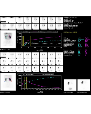
Renal scintigraphy is a highly sensitive nuclear medicine imaging technique that evaluates renal structure, blood flow, and function using radiopharmaceuticals such as Tc-99m DTPA, Tc-99m MAG3, and Tc-99m DMSA. Unlike anatomical imaging methods, renal scintigraphy provides dynamic physiological data, making it indispensable for assessing differential renal function, drainage patterns, and parenchymal integrity. It is safe, minimally invasive, and suitable for both adults and children.
How Renal Scintigraphy Works
Depending on the tracer used, renal scintigraphy assesses distinct functional parameters—DTPA reflects glomerular filtration, MAG3 measures tubular secretion and drainage, while DMSA provides high-resolution cortical imaging. Functional data is acquired in real time, allowing accurate evaluation of renal perfusion, excretory kinetics, and urinary tract obstruction. The radiation dose is low, and radiotracers are rapidly cleared, making the procedure well tolerated even in pediatric populations.
Clinical Importance and Evidence-Based Utility
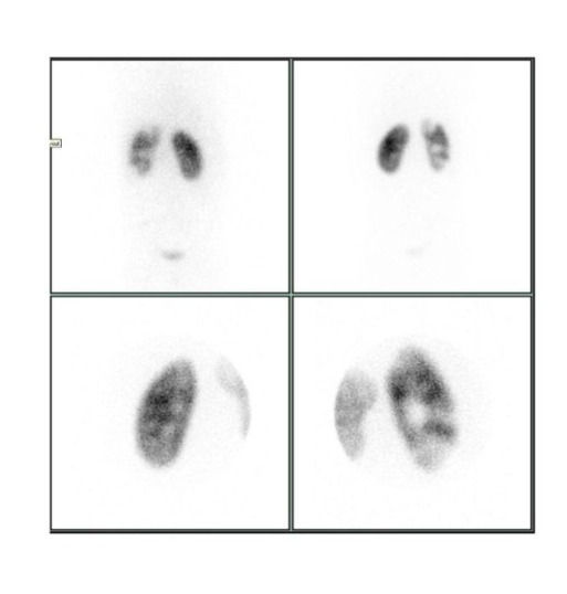
Renal scintigraphy plays a pivotal role in diagnosing obstructive uropathy, renovascular hypertension, pyelonephritis, congenital anomalies, and postoperative renal function. It guides decision-making for stenting, nephrostomy, surgical reconstruction, or nephrectomy by quantifying split renal function and identifying salvageable renal tissue. Research consistently supports its superiority in functional assessment compared to ultrasound or CT, particularly in early obstruction and differential kidney performance.
Key Clinical Indications for Renal Scintigraphy
Functional evaluation where anatomical imaging is insufficient
Suspected ureteric obstruction or impaired renal drainage
Differential renal function assessment before surgery
Evaluating renovascular hypertension with ACE-inhibitor scans
Acute or chronic pyelonephritis using DMSA imaging
Postoperative evaluation after pyeloplasty or ureteric repair
Congenital abnormalities such as PUJ obstruction or duplex kidneys
Assessing renal transplants for perfusion, viability, or rejection
Diagnostic Advantages and Clinical Benefits
Accurate functional insights for superior management decisions
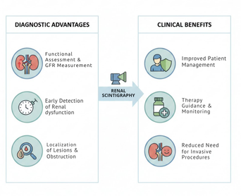
Provides real-time renal perfusion and excretion parameters
Differentiates obstructive from non-obstructive hydronephrosis
Quantifies split renal function with high reproducibility
Guides nephron-sparing decisions in adults and children
Helps evaluate medical therapy response in renovascular disease
Procedure, Safety, and Patient Workflow
A low-dose, well-tolerated imaging technique with high diagnostic value
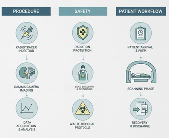
Performed with intravenous radiotracer injection
Dynamic imaging for MAG3 or DTPA to assess function and drainage
Optional diuretic (Lasix) or ACE-inhibitor augmentation when indicated
No need for sedation in most cases due to short, comfortable procedure
Minimal radiation exposure suitable for pediatric patients
Why Choose Us
Excellence in Advanced Nuclear Medicine, Precision Imaging, and Targeted Therapies
Precision-driven nuclear imaging ensuring unmatched diagnostic clarity and confidence
Advanced theranostics delivering targeted, personalized cancer treatment solutions
Expert-led clinical care prioritizing safety, accuracy, and patient well-being
Comprehensive services covering oncology, cardiology, and functional imaging needs
Decades of experience supported by global research, leadership, and innovation

Our Expert Doctors

Dr. Gayana Shankaramurthy
