
Imaging for Avascular Necrosis
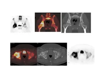
Understanding Imaging in Avascular Necrosis
Avascular necrosis (AVN), also known as osteonecrosis, results from compromised blood supply leading to bone ischemia and structural collapse. Early diagnosis is critical to prevent joint destruction, and imaging plays the central role in identifying disease long before symptoms become severe. Modern imaging modalities detect ischemic marrow changes, subchondral fractures, and cartilage involvement with high diagnostic precision, allowing timely intervention and preservation of joint function.
MRI as the Gold Standard for Early Detections
MRI remains the most sensitive and specific imaging technique for diagnosing AVN, capable of detecting bone marrow edema, early infarcts, and subtle subchondral changes before radiographs become abnormal. It provides excellent contrast resolution, detailed mapping of lesion extent, and accurate staging based on marrow signal characteristics. With its ability to reveal early pre-collapse disease, MRI is essential for treatment planning, prognosis, and monitoring of therapeutic response.
Complementary Role of X-ray, CT, and Nuclear Medicine
While plain radiographs help stage advanced AVN and identify structural deformity, they often appear normal in early disease. CT provides superior visualization of subchondral fractures, collapse patterns, and bone trabecular integrity, making it valuable in surgical planning. Nuclear medicine techniques such as bone scintigraphy can detect early perfusion deficits, particularly in multifocal AVN, and remain useful when MRI is contraindicated. Together, these modalities ensure a comprehensive evaluation of disease extent and joint viability.
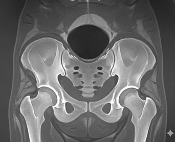
MRI Evaluation in Avascular Necrosis
High-resolution assessment for early diagnosis and treatment planning
Detects early ischemic changes before radiographs show abnormalities
Identifies classic “double-line sign” and subchondral crescent fractures
Maps lesion size, location, and cartilage involvement
Helps stage AVN (Ficat, ARCO) to guide treatment decisions
Monitors healing or progression after core decompression or biologics
Essential for evaluating bilateral or multifocal disease in high-risk patients
CT and X-ray for Structural Integrity and Collapse Evaluation
Precise visualization of bone architecture, subchondral defects, and joint preservation potential
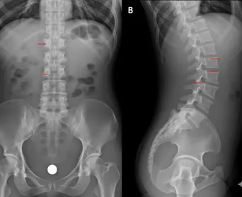
X-rays detect sclerosis, cysts, collapse, and joint space narrowing
CT provides detailed 3D assessment of subchondral fractures
Helps determine candidacy for joint-preserving vs joint-replacement procedures
Useful in cases with metallic implants or MRI contraindications
Supports surgical planning for osteotomies and resurfacing techniques
Nuclear Medicine and Functional Bone Assessment
Physiologic imaging to evaluate bone perfusion and disease extent
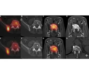
Bone scintigraphy detects early perfusion defects in ischemic bone
Useful for multifocal AVN evaluation in corticosteroid or alcohol-related cases
Helps assess viability of femoral head before joint-preserving surgery
SPECT-CT improves localization and characterization of necrotic areas
Beneficial when MRI is unavailable or inconclusive
Why Choose Us
Excellence in Advanced Nuclear Medicine, Precision Imaging, and Targeted Therapies
Precision-driven nuclear imaging ensuring unmatched diagnostic clarity and confidence
Advanced theranostics delivering targeted, personalized cancer treatment solutions
Expert-led clinical care prioritizing safety, accuracy, and patient well-being
Comprehensive services covering oncology, cardiology, and functional imaging needs
Decades of experience supported by global research, leadership, and innovation

Our Expert Doctors

Dr. Gayana Shankaramurthy
