
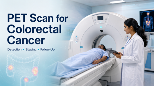
A PET scan can provide important information when colorectal cancer is suspected to have spread, persisted or returned after treatment. By revealing areas of increased metabolic activity, PET/CT may help identify active disease that requires further assessment.
Dr. Gayana Shankaramurthy, a Nuclear Medicine and Theranostics Specialist in Bangalore, emphasises that PET/CT findings are most meaningful when correlated with pathology, CEA trends, previous scans and treatment history. This complete clinical review helps determine whether the findings could influence the next care decision.
A PET Scan for Colorectal Cancer may be recommended for rising CEA levels, uncertain CT or MRI findings, suspected recurrence, or the evaluation of liver and distant metastases. It complements colonoscopy, biopsy and structural imaging, helping the care team plan the appropriate investigation or treatment pathway.
Is PET/CT required for every person diagnosed with colorectal cancer? Let’s discuss the situations in which it may provide useful additional information.
When Is a PET Scan Used for Colorectal Cancer?
PET/CT usually isn’t the first test to diagnose a colorectal tumour. Colonoscopy and biopsy are needed, these tests examine the bowel lining and confirm whether suspicious tissue is cancerous.
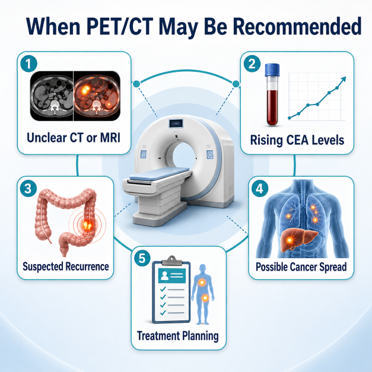
A PET scan is considered when:
* CT or MRI findings are unclear
* There’s a suspicion cancer has returned after treatment
* CEA levels are rising without a clear explanation
* Liver or lung metastases need to be assessed for possible treatment
* The care team needs to look for disease outside a known area
* Additional staging information could change the treatment or surgical plan
* Response to selected treatments needs a further evaluation
The decision is individual. Recent surgery, chemo, or radiation can affect how the tracer gets taken up and therefore influence the timing and interpretation of the examination.
The right scan, done at the right time, can answer a specific clinical question. Reach out for a careful review of your existing reports and imaging.
So what actually lets a PET scan pick up on active disease? Here’s the science, in plain terms.
How a PET Scan Detects Colorectal Cancer Cells
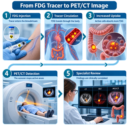
- FDG Injection: A small amount of FDG is injected into a vein.
- Tracer Absorption: The tracer circulates through the body and may accumulate in cells with increased metabolic activity.
- PET Imaging: The PET scanner detects signals emitted by the tracer and highlights areas of increased uptake.
- CT Localisation: CT images show the precise anatomical location of the highlighted areas.
- Specialist Interpretation: A nuclear medicine physician compares the findings with previous scans, pathology, CEA levels and treatment history.
Increased FDG uptake does not always indicate cancer because infection, inflammation and healing tissue can also appear active. Small or less metabolically active tumours may also be difficult to detect.
PET Scan for Detecting Liver and Distant Metastasis
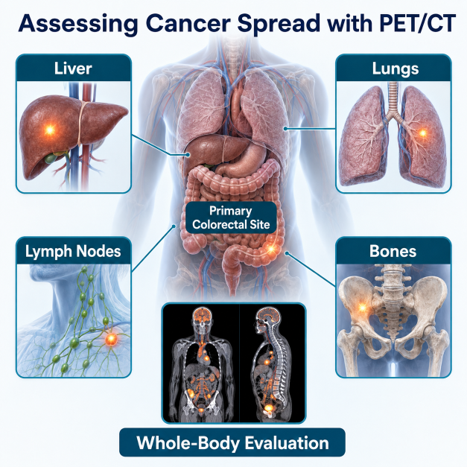
- Liver Assessment: Detects metabolically active lesions that may represent liver metastases.
- Whole-Body Evaluation: Examines multiple regions for signs of cancer spread in a single scan.
- Extrahepatic Disease Detection: May reveal disease outside the liver that was unclear on earlier imaging.
- Treatment Planning: Helps determine whether surgery or liver-directed treatment may remain suitable.
- Combined Interpretation: PET findings are reviewed alongside contrast-enhanced CT, liver MRI, pathology and clinical records.
Very small lesions and certain tumour types may show limited FDG uptake. Therefore, PET/CT complements rather than replaces detailed liver imaging.
PET Scan for Rising CEA Levels After Treatment
CEA is a tumour marker monitored in some patients after colorectal cancer treatment. A consistent rise may require further investigation, but it does not confirm cancer recurrence on its own.
- Reviews CEA Trends: Doctors assess changes across multiple blood tests rather than relying on a single result.
- Investigates Possible Recurrence: PET/CT may help locate metabolically active disease when recurrence is suspected.
- Clarifies Uncertain Findings: It can provide additional information when CT, MRI or colonoscopy results are inconclusive.
- Examines the Whole Body: The scan may identify active disease in the liver, lungs, lymph nodes, bones or other areas.
- Supports Further Planning: Findings may guide additional imaging, biopsy or treatment decisions.
Dr. Gayana Shankaramurthy, explains, “A rising CEA level should be interpreted alongside previous values, symptoms, treatment history and imaging findings. PET/CT is most useful when it addresses a specific clinical concern rather than being assessed in isolation.”
Distinguishing Scar Tissue from Cancer Recurrence
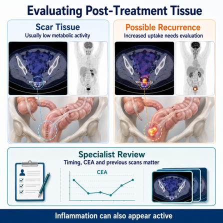
Surgery and radiation therapy can leave scar tissue, thickening or altered anatomy that remains visible on CT or MRI. PET/CT adds metabolic information to help assess whether the tissue appears inactive or requires further investigation.
- Assesses Metabolic Activity: Scar tissue generally shows little FDG uptake, while active cancer may absorb more tracer.
- Clarifies Structural Changes: PET/CT can provide additional information when CT or MRI shows a persistent mass or thickened area.
- Compares Previous Scans: Changes in size and metabolic activity over time can support more accurate interpretation.
- Considers Treatment Timing: Recent surgery or radiation may cause inflammation that appears active on PET.
- Guides Further Evaluation: Uncertain findings may require follow-up imaging, colonoscopy or biopsy.
PET/CT cannot always distinguish recurrence from inflammation on its own. Findings should be interpreted with the treatment timeline, CEA levels, pathology and previous imaging.
PET Scan vs Colonoscopy and Other Diagnostic Tests
| Test | What it primarily shows | Common role |
| Colonoscopy | The inner surface of the colon and rectum | Detecting tumours or polyps and collecting a biopsy |
| CT scan | Detailed structural images of the chest, abdomen and pelvis | Initial staging, treatment planning and follow-up |
| MRI | High-detail soft-tissue anatomy | Local rectal staging and detailed liver assessment |
| PET/CT | Metabolic activity combined with anatomical location | Investigating recurrence, uncertain findings or distant spread |
| CEA blood test | Changes in a tumour-marker level | Monitoring trends after treatment in suitable patients |
How to Prepare for a PET Scan
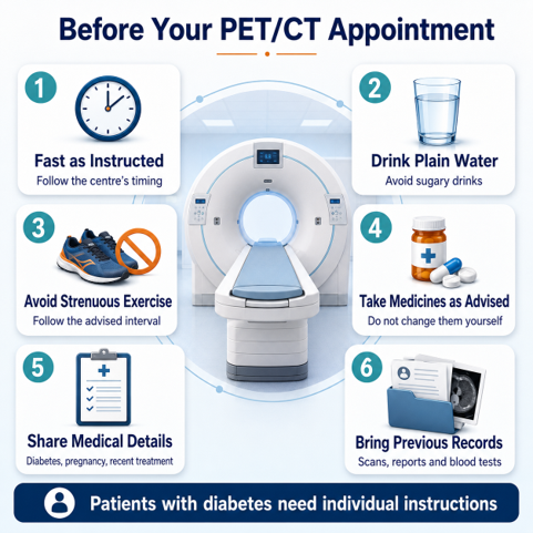
Patients should follow the imaging centre’s instructions because preparation can vary. Typical guidance includes:
- Avoid food for the stated number of hours before the scan
- Drink plain water unless fluid intake has been restricted
- Avoid sugary drinks, chewing gum and caloric beverages
- Avoid strenuous exercise for about 24 hours if instructed
- Continue medicines unless the care team advises otherwise
- Inform the team about diabetes, pregnancy or breastfeeding
- Report recent infections, procedures and cancer treatments
- Carry previous scans, reports, pathology results and CEA records
- Arrive on time because the radiotracer has a short usable period
Diabetic patients need individual instructions because blood glucose and insulin levels can influence FDG distribution. They should not change insulin, tablets or meal timings without professional guidance.
Is a PET Scan Safe for Colorectal Cancer Patients?
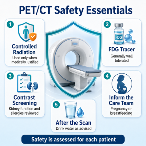
Generally speaking, yes — PET/CT is considered safe when medically indicated. The radiation dose is controlled, and the expected diagnostic benefit is weighed carefully against any potential risk.
- Controlled Radiation Exposure: Radiation comes from the FDG radiotracer and the CT component of the examination.
- Generally Well-Tolerated Tracer: Significant allergic reactions to FDG are rare.
- Contrast Assessment: If CT contrast is required, kidney function, allergies and previous contrast reactions may be reviewed.
- Pregnancy and Breastfeeding: Patients need to flag this with the imaging team beforehand — pregnant, possibly pregnant, or breastfeeding all matter here.
- After-Scan Care: Drinking water and passing urine regularly can help remove the tracer from the body.
Dr. Gayana Shankaramurthy highlights, “Patient safety depends on appropriate referral, correct preparation, optimised imaging, and experienced interpretation — these measures help obtain useful information while reducing unnecessary exposure or repeat scans.”
Frequently Asked Questions
Can a PET scan diagnose colorectal cancer?
Is PET/CT routinely used for initial colorectal cancer staging?
Can PET/CT detect colorectal cancer recurrence?
Can PET distinguish scar tissue from active cancer?
Which is better, colonoscopy or PET/CT?
Why must I fast before an FDG PET scan?
Does a negative PET scan rule out colorectal cancer?
References:
https://pmc.ncbi.nlm.nih.gov/articles/PMC2582503/
https://www.sciencedirect.com/science/article/pii/S0960740424001191
Disclaimer: The information shared in this content is for educational purposes and not for promotional use.
